RGUHS Nat. J. Pub. Heal. Sci Vol No: 17 Issue No: 4 pISSN:
Dear Authors,
We invite you to watch this comprehensive video guide on the process of submitting your article online. This video will provide you with step-by-step instructions to ensure a smooth and successful submission.
Thank you for your attention and cooperation.
1Dr. Tridib Nath Banerjee, Department of Prosthodontics, Dr. R Ahmed Dental College & Hospital, Kolkata, West Bengal, India.
2Department of Prosthodontics, Dr. R Ahmed Dental College & Hospital, Kolkata, West Bengal, India
3Department of Prosthodontics, Dr. R Ahmed Dental College & Hospital, Kolkata, West Bengal, India
4Department of Prosthodontics, Dr. R Ahmed Dental College & Hospital, Kolkata, West Bengal, India
*Corresponding Author:
Dr. Tridib Nath Banerjee, Department of Prosthodontics, Dr. R Ahmed Dental College & Hospital, Kolkata, West Bengal, India., Email: tridibbanerjee81@gmail.com
Abstract
The curve of Spee, an anteroposterior curvature of the plane of occlusion, has long intrigued dental professionals. This review delves into its historical roots, development, and various influencing factors like age and sex, thus shedding light on its pivotal role in prosthodontics. In natural dentition, this curvature supports balanced interactions between anterior teeth, posterior teeth, and the condylar path. During the fabrication of complete dentures, the anteroposterior curve is incorporated to achieve occlusal balance, facilitate protrusive disocclusion, prevent interferences, and optimize masticatory force distribution. Understanding and managing this curvature is therefore crucial for achieving optimal outcomes in complete dentures and full mouth rehabilitations. Future research should explore individual variability, interactions with other occlusal factors, and advanced measurement and incorporation techniques.
Keywords
Downloads
-
1FullTextPDF
Article
Introduction
In edentulous patients undergoing dental restoration, determining the optimal occlusal plane is paramount, as it dictates the ideal positioning of the prosthetic teeth for both function and aesthetics.1,2 Proper management of the occlusal plane is essential when designing restorations in long-span posterior edentulous areas or when incorporating restorations into an existing dentition with rotated, tipped, or extruded teeth. It necessitates careful consideration of potential excursive interferences arising from the altered occlusal plane, deviating from the ideal harmonious interaction connecting the anterior teeth and condylar guidance.3 The contrastive analysis of the above-mentioned relationship will be a helpful guide during the positioning of artificial teeth for complete denture prosthesis and the determination of the occlusal plane in full mouth reconstruction, leading to optimum functional occlusion.
Historical Overview
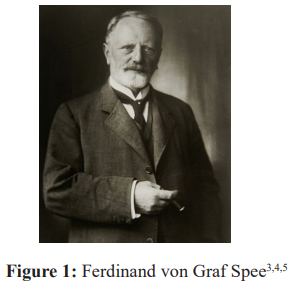
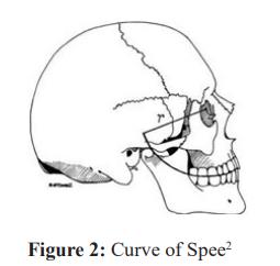
The curve of Spee was reported first by Ferdinand von Graf Spee (Figure 1) in 1890 as an anteroposterior anatomic curve touching the tip of cusps of anterior teeth, the buccal cusps of premolar and molar teeth, and continuing posteriorly to the end at the frontal most part of mandibular condyle (Figure 2).3 This anteroposterior curve, present in the occlusal plane of natural dentition, promotes harmonious interaction between anterior tooth guidance, posterior teeth and condylar path. It is introduced during the fabrication of complete denture to attain occlusal balance.
In 1890, Ferdinand von Graf Spee pioneered the concept of the “Curve of Spee” by analysing worn human teeth.4 He proposed that this curved occlusal alignment, tangential to a cylinder touching the condyle, second molar, and incisors, represents the ideal arrangement for balanced jaw function. This hypothetical cylinder, encompassing the occlusal line, has a radius which was estimated by Spee to be 6.5-7.0 cm, with its centre residing in the axial plane at the level of the mid-orbit (2.5 inches). Spee noted the possibility of locating the centre of the curvature by calculation using a compass.4 He postulated that the curve served as a critical design feature, ensuring continuous tooth contact during both forward and backward jaw movements crucial for efficient chewing. Recognizing the functional importance of the curvature, Spee advocated for its incorporation into denture design. This aimed to improve chewing efficiency and mitigate potential lever effects caused by uneven forces during mastication. Analysis of the curvature may assist clinicians in determining the plane of occlusion in sagittal axis. The curve of Spee, present in both the maxillary and mandibular arches, holds significant value in prosthetic and orthodontic reconstruction. Studies have consistently shown that optimal sagittal organization of teeth, as guided by this curvature, directly impacts the functional stability and success of complete dentures and implant-supported restorations. Conversely, deviations from this ideal alignment can lead to excessive forces, uneven distribution of pressure, and ultimately, restoration failure.
Evolution of Curve of Spee
The curve of Spee (Figure 2) represents the anatomical curvature of the plane of occlusion, when viewed from the lateral aspect. It starts at the tip of the lower canine, tracing along the buccal cusp tips of the premolar and molar teeth, follows the anterior border of the ramus of mandible, and finally connects to the anterior most part of the mandibular condyle.6
In 1926, an anthropological study by George Monson advocated the idea as suggested by Spee. He further described it as a sphere in three-dimensional plane, passing through the edges of the incisors and chewing surfaces of the teeth in the mandibular arch. Monson is credited with introducing the now well-established ‘curve of Spee’ of four inch radius.7 Okeson JP stated while examining the dental arches from lateral view, that if a virtual line is drawn through the tips of buccal cusps of posterior teeth (molars and premolars), a curved line following the plane of occlusion will be set up that is convex and concave for the maxillary and the mandibular arches, respectively.8 There is an ideal match between these convex and concave lines when dental arches are placed in occlusion. This curvature of the dental arches is referred to as the ‘curve of Spee’.
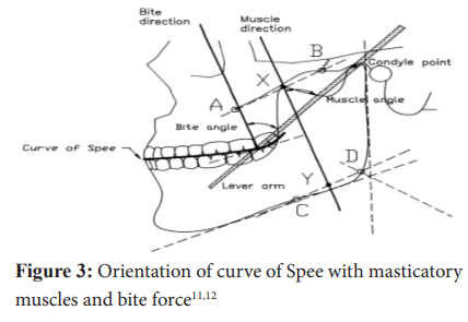
Peter E Dawson mentioned the curve of Spee as the anteroposterior curvature of the occlusal plane of the lower teeth. It commences at the cusp tip of lower canine and follows the tips of the buccal cusps of premolars and molars, continuing along the anterior border of the mandibular ramus until the anterior most portion of the mandibular condyle.9 According to Sicher, the formation of Spee’s curve results from the coincidence of the longitudinal axis of each individually attached and movable tooth with the direction of the resultant masticatory force at that point. This curved alignment redirects the downward biting force from jaw muscles into an angled force acting on each tooth (Figure 3). The resultant oblique force distribution reduces stress on individual teeth and supports their long-term health. The tilting of chewing forces on the molars in forward direction, together with their positional arrangement, plays a crucial role in creating the curved occlusal plane.10 Kleinberg further emphasized that the curve of Spee is necessary for enabling protrusive contact of the incisor teeth without causing interferences from the posterior teeth.11
Age and Gender Influence on Curve of Spee
Farella M and Martina R investigated the correlation between the curve of Spee (arch-like structure of the mandibular teeth) and skeletal facial features. The results indicated a significant presence of the curve in short-face individuals and less prominent in long-face individuals. This curvature is independent of the gender, age and amount of curvature in adults.13 Marshall S et al., studied the curve of Spee depth in the study models of subjects aged between 4 and 26 years and concluded that the curve of Spee initially develops as a consequence of permanent mandibular first molar and incisor eruption. This curvature maintains a consistent depth until the eruption of permanent second molars, at which point it further deepens. During adolescence, the curve exhibits a slight decrease in depth and stabilizes in early adulthood. Notably, these studies found that the differences in maximum curve of Spee depth between the right and left sides of the mandible, as well as between genders, were statistically nonsignificant.14
A study by Ferrario VF and Miani A found that the occlusal curvature of the mandibular arch was not significantly influenced by gender. The radii of Spee’s curve in the right and left molar area were about 105 mm in men and 100 mm in women. Within genders, the mean radii of left and right curves of Spee were identical.15 Ferrario VF and Sforza C in a study found that the right and left sides in males showed different concavities, with the right-hand side being flatter than the left. The study observed statistically notable differences in curve depths between the mandibular left and right sides, independent of gender.16 Hui Xu et al., reported that the curve of Spee had a mean radius of approximately 106.4 mm in the maxillary arch and 83.4 mm in the mandibular arch.17 The curve of Spee was not impacted by the gender of the subjects investigated. Braun ML & Schmidt WG in a cephalometric study found that the Spee’s curve was identical in Angle Class I and Class II Division I occlusions. They further suggested that its shape was similar in both males and females.18 Ferrario et al., demonstrated that in the mandibular dental arches, the radii of the curves of Monson, Spee and Wilson in the three-dimensional plane increased by approximately 20 mm during the transition from adolescence to early adulthood (from 80 mm at 12 to 14 years of age to 101 mm at 20 years of age). A high degree of symmetry was also observed between the radii of the left and right curves of Spee.19 The research conducted by Se-Hwan Cheon et al., revealed a positive association between the mandibular and maxillary curves of Spee. Sex was not found to be a significant predictor in the measurement of this curvature.20
Significance in Prosthodontics and Occlusion
According to Williamson, the rotated, tipped and extruded teeth may pathologically alter the curve of Spee. Replicating a modified occlusal plane during dental restoration can introduce interferences in the posterior region during protrusion, disrupting smooth jaw movements. This disruption can trigger abnormal activity in the masseter and temporalis muscles, potentially leading to muscle dysfunction and pain.21 Little AP stated that the projection of the curve of Spee toward the condyle crosses the long fibres of the masseter muscle at right angles. The resultant pull of the masseter muscle acts perpendicular to the curve of Spee. He further commented that the curves are extremely important for uniform distribution of applied stresses of mastication without excessive traumatic occlusion.22 According to Willand L, the occlusal plane guide and former allows the setting of posterior teeth resulting in development and preservation of the mesiodistal and buccolingual curves. The buccal cusps deviate from the occlusal plane in a continuous upward direction, forming a curve, while the lingual cusps remain aligned with the plane's reference level.23
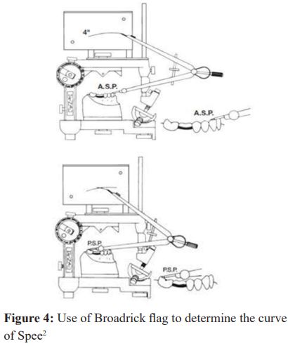
Hobo and Takayama recommended creating a curvature with a radius of 180 mm on the mandibular teeth, with a maximum depth of 3 mm at the level of the mesiobuccal cusp of the second molar, for full mouth rehabilitation of the occlusal plane.24 Lynch CD and McConnell RJ recommended the use of the Broadrick flag (Figure 4), that empowers dentists to create natural-looking and functional dentures and restorations by facilitating precise curve of Spee determination.2 Utilizing a standard 4-inch curve in patients presenting with a retrognathic mandible can lead to an undesirable flattened posterior occlusal plane. This flattened plane can subsequently induce posterior protrusive interferences, hindering smooth jaw movements during biting. Hence, an existence of a Class II skeletal relationship requires a 3.75-inch curve. On the contrary, the standard 4-inch curve might not be suitable for patients with a skeletal Class III, as it creates an excessive posterior slope, resulting in posterior protrusive interferences with biting. The application of the Broadrick flag (Figure 4) helps in the determination of an acceptable curve of occlusion for individual dentate patient. A 5-inch radius would be ideal in this situation. Starcke EN recommended the Ortho Plane analyzer from Ivoclar as part of the biogenic prosthetic technique, to assist in forming the occlusal rims and serve as a guide for arranging denture teeth along a predetermined curve of Spee.25 KaVo EWL as an accessory of the PROTAR articulator system also provided a curved template of 125 mm radius for setting denture teeth. According to Dawson, the curve of Spee results from variations in axial positioning of the lower teeth when last molar tilts forward at the greatest angle and the forward tooth is at least angle. The curved occlusal plane allows for the posterior teeth to disengage (disocclusion) during forward jaw movement (protrusive) through a coordinated interplay between the condylar guidance and incisal guidance.9 Kleinberg added that curve of Spee is required to prevent interferences in posterior teeth by allowing protrusive contact of anterior teeth.1
Discussion
The functional significance of the curve has been explored by several researchers. Spee proposed the curvature as the optimal model for maintaining tooth contact during mandibular protrusive and retractive movements while chewing. The sagittal arrangement of teeth is controlled by the articular eminence angle, incisal overlap, molar cusp height, and posterior contact area.18 These factors, along with the curvature, contribute to balanced chewing function. Recent research suggests that the curve of Spee elevates the crush-shear ratio between posterior teeth, playing a biomechanistic role in food processing and thereby enhancing chewing efficiency.26 Analysis of this occlusal curvature can help dentists in establishing optimal sagittal occlusal relationships in restorations, using both maxillary and mandibular curves as references.18
Conclusion
The curve of Spee is not merely an anatomical curiosity but plays a crucial role in functional occlusion and mastication. This review explored the historical background, evolution, and various influences of this occlusal curvature, highlighting its importance in prosthetic dentistry and occlusion. The curve facilitates protrusive disocclusion of posterior teeth, preventing interference during forward jaw movement. It optimizes the distribution of masticatory forces, reducing stress on individual teeth and muscles. Understanding and managing the curve is essential for restoring optimal occlusal function in complete denture prostheses and full mouth rehabilitations. By integrating this curvature into treatment planning and execution, prosthodontists can achieve more natural, stable, and functional occlusal outcomes for their patients.
Conflict of Interest
Nil
Supporting File
References
- Shigli K, Chetal BR, Jabade J. Validity of soft tissue land marks in determining the occlusal plane. J Indian Prosthodont Soc 2005;5(3):39-45.
- Lynch CD, McConnell RJ. Prosthodontic management of the curve of Spee: Use of the Broadrick flag. J Prosthet Dent 2002;87(6):593-97.
- Von Spee FG. The condylar path of the mandible in the glenoid fossa. Arch Anat Physiol 1890;16: 285-294.
- Spee FG, Biedenback MA, Hotz M, et al. The gliding path of mandible along the skull. J Am Dent Assoc 1980;100(5):670-5.
- Hill MA. Ferdinand Graf Von Spee.jpg. [Internet]. Sydney: UNSW Embryology.
- The Glossary of Prosthodontic Terms: Ninth Edition. J Prosthet Dent 2017;117(5S):e1-e105.
- Monson GS. Applied mechanics to the theory of mandibular movements. Dent Cosmos 1932;74:1039-53.
- Okeson JP. Management of temporomandibular disorders and occlusion. 6th ed. St Louis: Mosby; 2008.
- Dawson PE. Functional occlusion from TMJ to smile design. Mosby: Elsevier; 2007.
- Sicher H, DuBrul EL. Sicher and DuBrul’s Oral Anatomy. 8th edition. St. Louis, MO: Ishiyaku EuroAmerica Inc.; 1988.
- Klineberg I. Occlusion: Principles and assessment. Oxford, United Kingdom: Buterworth-Heinemann; 1992.
- Osborn JW. Orientation of the masseter muscle and the curve of Spee in relation to crushing forces on the molar teeth of primates. Am J Phys Anthropol 1993;92(1):99-106.
- Farella M, Michelotti A, van Eijden TM, et al. The curve of Spee and craniofacial morphology: A multiple regression analysis. Eur J Oral Sci 2002;110(4):277-81.
- Marshall SD, Caspersen M, Hardinger RR, et al. Development of the curve of Spee. Am J Orthod Dentofacial Orthop 2008;134(3):344-52.
- Ferrario VF, Miani A. Mathematical definition of the curve of Spee in permanent healthy dentitions in man. Arch Oral Biol 1992;37(9):691-4.
- Ferrario VF, Sforza C, Miani A Jr. Statistical evaluation of Monson’s sphere in healthy permanent dentitions in man. Arch Oral Biol 1997;42(5): 365-9.
- Xu H, Suzuki T, Muronoi M, et al. An evaluation of the curve of Spee in the maxilla and mandible of human permanent healthy dentitions. J Prosthet Dent 2004;92(6):536-9.
- Braun ML, Schmidt WG. A cephalometric appraisal of the curve of Spee in Class I and Class II, Division I occlusions for males and females. Am J Orthod 1956;42(4):255-8.
- Ferrario VF, Sforza C, Poggio CE, et al. Three-dimensional dental arch curvature in human adolescents and adults. Am J Orthod Dentofacial Orthop 1999;115(4):401-5.
- Cheon SH, Park YH, Paik KS, et al. Relationship between the curve of Spee and dentofacial morphology evaluated with a 3-dimensional reconstruction method in Korean adults. Am J Orthod Dentofacial Orthop 2008;133(5):640.e7-14.
- Williamson EH, Lundquist DO. Anterior guidance: Its effect on electromyographic activity of the temporal and masseter muscles. J Prosthet Dent 1983;49(6):816-23.
- Little AP. Practical bio-engineering in full denture construction. JADA 1935;22(6):1016.
- Willand L. Occlusal plane guide and former. J Prosthet Dent 1969;22(6):671-8.
- S. Hobo S, Takayama H. Oral rehabilitation, clinical determination of occlusion. Carol Stream, Illinois: Quintessence Publishing Co. Inc; 1997.
- Starke EN. The history of articulators: A critical history of articulators based on geometric theories of mandibular movement-Part I. J Prosthodont 2002;11(2):134-46.
- Ash MM, Nelson SJ. Wheeler’ dental anatomy, physiology and occlusion. 8th Edition. Elsevier Science; 2003.
