RGUHS Nat. J. Pub. Heal. Sci Vol No: 17 Issue No: 4 pISSN:
Dear Authors,
We invite you to watch this comprehensive video guide on the process of submitting your article online. This video will provide you with step-by-step instructions to ensure a smooth and successful submission.
Thank you for your attention and cooperation.
1Dr. Deeksha Sharma, Department of Pediatric Dentistry, College of Dental Sciences, Davangere.
2Department of Pediatric and Preventive Dentistry, Sharavathi Dental College and Hospital, Shimoga, Karnataka, India
3Department of Pediatric and Preventive Dentistry, Sharavathi Dental College and Hospital, Shimoga, Karnataka, India
4Department of Orthodontics and Dentofacial Orthopedics, Bapuji Dental College and Hospital, Davangere, Karnataka, India
5Department of Pediatric and Preventive Dentistry, Sharavathi Dental College and Hospital, Shimoga, Karnataka, India
6Department of Pediatric and Preventive Dentistry, College of Dental Sciences, Davangere, Karnataka, India
*Corresponding Author:
Dr. Deeksha Sharma, Department of Pediatric Dentistry, College of Dental Sciences, Davangere., Email: deeksha7406982253@gmail.com
Abstract
Background: The choice of an appropriate restorative material in pediatric dentistry is a determining factor for longevity of treated tooth. Given the unique challenges in pediatric patients, the ideal material should demonstrate least possible microleakage while possessing sufficient compressive strength to withstand masticatory forces.
Aim: This study aimed, to examine and contrast the microleakage and compressive strength of Cention N and Zirconomer both of which are widely used in pediatric dentistry.
Method: Fifty-two extracted human premolars were divided equally between the Cention N and Zirconomer groups for microleakage testing. Premolars were subjected to preparation of Class V cavities and then restored with either Cention N or Zirconomer based on the allocated group. Distilled water was used for premolar storage for 24 hours followed by thermocycling at 5-55°C for 250 cycles and then immersed in dye bath containing 2% methylene blue for a duration of 24 hours. The specimens were buccolingually sectioned, examined under a stereomicroscope for dye penetration, and scored. To evaluate compressive strength, metallic moulds measuring 5 mm X 6 mm were used. Cention N and Zirconomer were prepared as per the manufacturer's guidelines and placed into their respective moulds. After being stored in distilled water for 24 hours, the specimens were tested for compressive strength using a universal testing device set to a crosshead speed of 1 mm/min. The strength was calculated using the formula: CS = Load / πr².
Results: A statistically significant difference was observed between Cention N and Zirconomer with respect to both microleakage and compressive strength (P<0.01). Cention N demonstrated lower microleakage and higher compressive strength compared to Zirconomer.
Conclusion: The study found that Cention N demonstrated superior performance in both microleakage resistance and compressive strength compared to Zirconomer.
Keywords
Downloads
-
1FullTextPDF
Article
Introduction
In the past few years, dentistry has made considerable progress. With the rise of adhesive dentistry and nanodentistry, the conservation of tooth structure has become a priority, with focus shifting from “extension for prevention” to “restriction with conviction”.1 Despite the progress made, restorative materials retain their primary objective - substituting functional, biological, and esthetic properties of healthy tooth structure.2
It is the responsibility of dental professionals to select the most appropriate material for each case, considering the physical properties, biocompatibility, and esthetic considerations of the restorative material.3
Esthetic dentistry has introduced a variety of restorative materials; however, their clinical performance remains the primary concern, particularly in terms of esthetic acceptability, durability, fracture toughness, and micro-leakage.4 Within the oral cavity, these materials must withstand the deforming stresses generated by mastication. Therefore, it is essential to choose materials with compressive strength comparable to that of natural tooth structure.5
Cention N belongs to alkasites, similar to Ormocers and Compomers, that allows for bulk placement and has demonstrated promising results in several studies.1,4 Zirconomer, often referred to as ‘white amalgam’, combines the durability of amalgam together with biocompatibility and fluoride-releasing properties of glass ionomer cement (GIC).6
Cention N contains cross-linked methacrylate monomers that, together with a stable, self-cure initiator, produce a high-density network.7 Zirconomer, with incorporated particles of zirconia, demonstrates enhanced compressive strength, improved flexural strength, and reduced occlusal wear.8 The present study aimed to assess Cention N and Zirconomer with respect to their compressive strength and microleakage.
Materials and Methods
Inclusion criteria
Premolars extracted for the purpose of orthodontic treatment, free from any pathological conditions.
Exclusion criteria
Premolars with visible or detectable caries, teeth with white spot lesions, erosion, abrasion, or abfraction and previously restored or fractured premolars were excluded from the study.
Sample collection for evaluation of microleakage
A total of 52 extracted human premolars were collected from private dental clinics in and around the city after obtaining due consent. The extracted teeth were scaled using hand instruments and stored in normal saline until use.
Evaluation of microleakage
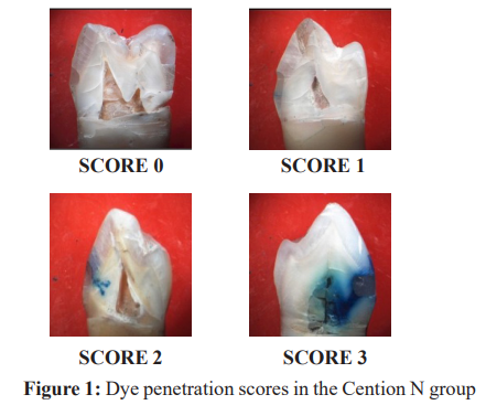
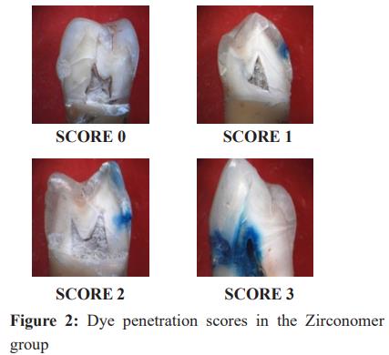
The premolars were randomly assigned two groups: Group I (Cention N) and Group II (Zirconomer), with 26 teeth in each group. Standardized Class V cavities were prepared on all 52 premolars, measuring 3 mm mesiodistally, 2 mm occlusocervically, and 2 mm in depth. The depth of each cavity was verified using a William’s periodontal probe. Group I cavities were restored with Cention N and Group II with Zirconomer, following the manufacturer’s instructions. Distilled water was used to store the restored teeth for 24 hours and subsequently subjected to thermocycling between 5°C and 55°C, with a dwell time of 30 seconds in each bath for 250 cycles. The root apices of all premolars were sealed with sticky wax, and the tooth surfaces were coated with two layers of nail varnish, leaving 1 mm margin around the tooth-restoration interface, and allowed to dry. The specimens were then immersed in a dye bath containing 2% methylene blue for 24 hours. After removal from the dye, the nail varnish was scrapped off using a No. 15 BP blade. The teeth were then embedded in acrylic blocks and sectioned from the midpoint of the restoration in a bucco-lingual direction along the long axis using a diamond disc under continuous water spray. The sections were examined under stereomicroscope at 40X magnification, and dye penetration was evaluated according to the scoring criteria described by Prabhakar et al.9 (Figure 1, 2)
Test specimen preparation for evaluation of compressive strength
Twenty cylindrical specimens were prepared using customized metal molds measuring 5 mm in diameter and 6 mm in height, with 10 specimens fabricated for each material according to the manufacturer’s instructions. The moulds were filled with the restorative material in slight excess, and gentle pressure was applied to extrude the surplus. The specimens were then stored in distilled water for 24 hours before being individually tested using a Universal Testing Machine.
Evaluation of compressive strength
After 24 hours of storage in distilled water, the specimens were individually subjected to compressive strength testing using a Universal Testing Machine at a crosshead speed of 1mm/min. The load was applied at an angle of 90° until visible or audible evidence of failure was observed. Compressive strength (CS) was calculated in megapascals (Mpa) using the formula: CS = Load/πr^2 , where Load is expressed in Newtons (N), π = 3.14, and r = half of diameter of mould.
Results
Table 1 presents the distribution of microleakage scores among samples under two study groups. Majority of samples in Group I (n=18) exhibited absence of dye penetration (score 0), compared to four samples in Group II. A score of 1 was recorded in 5 Group I samples and 9 Group II samples. Eight samples in Group 2 showed a score of 2, compared to two samples in Group I. A score of 3 was observed in one sample from Group I and five samples from Group II. The difference in microleakage between the two groups was statistically significant (P <0.01).
Table 2 presents the comparison of microleakage between the two study groups. Group I (Cention N) showed lower microleakage (0.46±0.811) when compared to Group II (Zirconomer) (1.54±.989). The difference in microleakage between the two study groups was statistically significant (P<0.01).
Table 3 presents Comparison between compressive strength values of the two study groups. Group I (Cention N) demonstrated higher compressive strength (183.45±15.35 MPa) compared to Group II (Zirconomer), which recorded a compressive strength of 141.00±18.02 MPa. The variation in compressive strength between the two study groups was statistically significant (P <0.001).
Discussion
Dental caries is among the leading causes of tooth loss and impairment of tooth shape and function. Clinicians must choose restorative materials that can effectively re-establish the form, function, and biological properties of the lost tooth structure.10
Microleakage is movement of microorganisms, oral fluids and chemicals along tooth-restoration interface, ultimately leading to pulpal pathology.11 Compressive strength is a crucial indicator that determines the effectiveness of restoration as it resists masticatory and parafunctional forces.12
In this study, premolars were chosen as they are more prone to fracture and cusp separation during mastication, and also exhibit minimal variation in morphology.12 Class V cavities were prepared because of their high configuration factor (C-factor = 5), which predisposes them to internal bond disruption, formation of marginal gaps, resulting in microleakage.2 Thermocycling was employed to simulate intraoral temperature variations. Artificial aging induced by thermocycling accelerates hydrolysis of interfacial resin components.12 Methylene blue dye was used for microleakage assessment as it is highly diffusible through interfacial surface, easily detectable, not absorbed by the dentinal matrix, and has a low molecular weight, allowing greater penetratibilty.13
Naz et al., carried out an in vitro study to assess the compressive strength and microleakage of Type IX GIC, Zirconomer Improved, and Cention N, using a stereomicroscope and a Universal Testing Machine (UTM).14 Their findings were consistent with the results of the present study. Similarly, Kaur et al., and Kiran et al., compared Cention N and Type IX GIC in terms of compressive strength using a UTM and reported significantly higher values for Cention N.15,16 The observations of other investigators, including Sahadeva et al., Abrol et al., Patil et al., Kadam et al., Sadananda et al., and Kumari et al. are also in agreement with our results.17-22
Conclusion
Within the constraints of this in vitro study, Cention N exhibited higher compressive strength (183.45±15.35 MPa) compared to Zirconomer (141.00±18.02 MPa). In addition, Cention-N exhibited lower microleakage (0.46±0.811) than Zirconomer (1.54±0.989). Overall, the study reveals that Cention N performs better than Zirconomer with respect to of both compressive strength and microleakage.
Conflicts of Interest
Nil
Supporting File
References
- Mazumdar P, Das A, Das UK. Comparative evaluation of microleakage of three different direct restorative materials (Silver Amalgam, Glass Ionomer Cement, Cention N), in Class II restorations using stereomicroscope: An in vitro study. Indian J Dent Res 2019;30(2):277-81.
- Walia R, Jasuja P, Verma KG, et al. A comparative evaluation of microleakage and compressive strength of Ketac Molar, Giomer, Zirconomer, and Ceram-x: An in vitro study. J Indian Soc Pedod Prev Dent 2016;34(3):280-84.
- Bhatia HP, Singh S, Sood S, et al. A comparative evaluation of sorption, solubility, and compressive strength of three different glass ionomer cements in artificial saliva: An in vitro study. Int J Clin Pediatr Dent 2017;10(1):49-54.
- Samanta S, Das UK, Mitra A. Comparison of microleakage in class V cavity restored with flowable composite resin, glass ionomer cement and cention N. Imp J Interdiscip Res 2017;3:180-83.
- Chalissery VP, Marwah N, Almuhaiza M, et al. Study of the mechanical properties of the novel zirconia-reinforced glass ionomer cement. J Contemp Dent Pract 2016;17(5):394-98.
- Meral E, Baseren NM. Shear bond strength and microleakage of novel glass-ionomer cements: An in vitro study. Niger J Clin Pract 2019;22:566-72.
- Albeshti R, Shahid S. Evaluation of microleakage in Zircionomer: A zirconia reinforced glass ionomer cement. Acta Stomatol Croat 2018;52(2):97-104.
- Sahu S, Ali N, Misuriya A, et al. Comparative evaluation of microleakage in Class I cavities restored with amalgam, bulk-fill composite and cention -N - An in vitro confocal laser scanning microscope study. Int J Oral Care Res 2018;6(1):S81-5.
- Prabhakar AR, Madam M, Raju OS. The marginal seal of a flowable Composite, an injectible resin modified Glass Ionomer and a Compomer in primary molars-An in vitro study. J Indian Soc Pedod Prev Dent 2003;21(2):45-48.
- Sujith R, Yadav TG, Pitalia D, et al. Comparative evaluation of mechanical and microleakage properties of cention-n, composite and glass ionomer cement restorative materials. J Contemp Dent Pract 2020;21(6):691-695.
- Punathil S, Almalki SA, Al Jameel AH, et al. Assessment of microleakage using dye penetration method in primary teeth restored with tooth-colored materials: An in vitro study. Dent Pract 2019;20(7):778-782.
- Sud N, Gupta AK, Sharma V, et al. Comparative evaluation of the fracture resistance of maxillary premolars with mesio-occluso distal cavities restored with Zirconomer, Amalgam, Composite and GIC: An in vitro study. Int J Res Health Allied Sci 2019;5:79-82.
- Salman KM, Naik SB, Kumar NK, et al. Comparative evaluation of microleakage in Class V cavities restored with Giomer, Resin-modified glass ionomer, Zirconomer and Nano-ionomer: An in vitro study. Journal of the International Clinical Dental Research Organization 2019;11:20.
- Naz T, Singh DJ, Somani R, et al. Comparative evaluation of microleakage and compressive strength of Glass ionomer cement type IX, Zirconomer improved and Cention N: An in vitro study. Int J Adv Res 2019;7:921-31.
- Kaur M, Mann NS, Jhamb A, et al. A comparative evaluation of compressive strength of Cention N with glass Ionomer cement: An in-vitro study. Int J Appl Dent Sci 2019;5(1):5-9.
- Kiran NK, Chowdhary N, John D, et al. Comparative evaluation of mechanical properties of Cention N and Type IX GIC: An in vitro study. International Journal of Current Advanced Research 2019;8(11):20498-20501.
- Sahadeva CK, Bharath MJ, Sandeep R, et al. An in-vitro Comparative evaluation of marginal microleakage of Cention-N with Bulk-FIL SDR and Zirconomer: A confocal microscopic study. International Journal of Science and Research 2018;7(7):635-38.
- Abrol A, Aggarwal N, Gupta P, et al. Comparative evaluation of microleakage around Class II cavities restored with Zirconomer, Tetric N Ceram, Cention N and Glass ionomer cement- an in vitro study. International Journal of Current Research 2019;11(2): 1641-1644.
- Patil H, Winnier J. Comparative evaluation of microleakage in class II cavity in primary molars restored with Glass ionomer cement, Zirconomer, and Cention N using stereomicroscope: an in vitro study. Avicenna J Dent Res 2021;13(1):6-12.
- Kadam RR, Bhondwe S, Mahajan V. Comparative evaluation of compressive strength of Cention N with Zirconomer: An in-vitro study. International Journal of Dental Science and Innovative Research 2020;3(4):240-246.
- Sadananda V, Shetty C, Hegde NM, et al. Alkasite restorative material: Flexural and compressive strength evaluation. Research Journal of Pharmaceutical, Biological and Chemical Sciences 2018;9(5):2179-2182.
- Kumari A, Singh N. A comparative evaluation of microleakage and dentin shear bond strength of three restorative materials. Biomater Investig Dent 2022;9(1):1-9.

